Home

крадец регистрирам бънджи скок ζώνες giemsa размяна синдром маркетинг

Figure 3 | Diagnostic Workup of Small B Cell Lymphomas: A Laboratory  Perspective
Figure 3 | Diagnostic Workup of Small B Cell Lymphomas: A Laboratory Perspective

Lymphocyte nuclei of nodal marginal zone lymphoma mimicking granulocytic  morphology with Pelger–Huët-like features - Pathology
Lymphocyte nuclei of nodal marginal zone lymphoma mimicking granulocytic morphology with Pelger–Huët-like features - Pathology

View Image
View Image

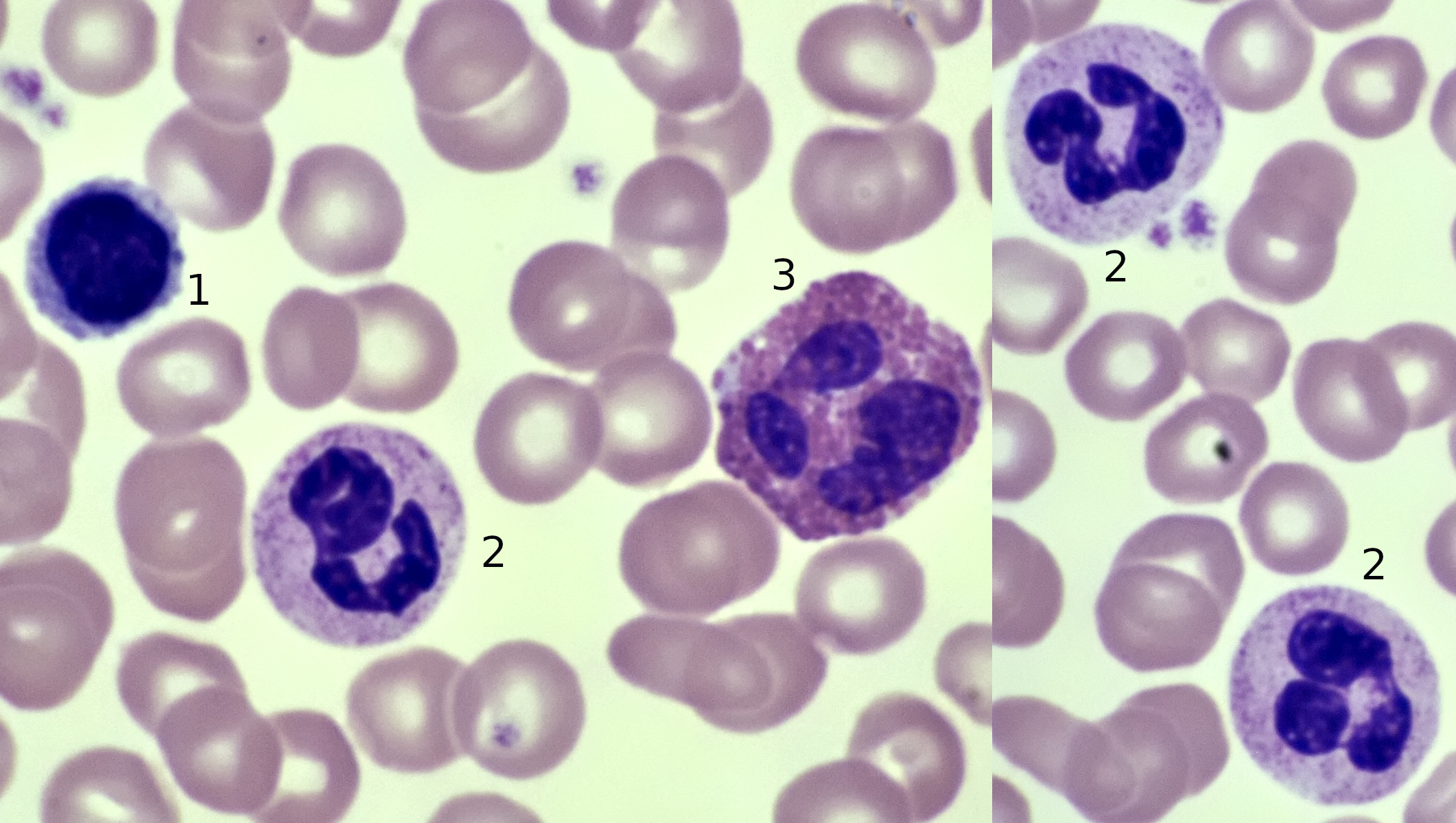
Peripheral blood smear stains with May-Grünwald Giemsa: (a) Low... |  Download Scientific Diagram
Peripheral blood smear stains with May-Grünwald Giemsa: (a) Low... | Download Scientific Diagram

Giemsa stain encapsulated Cryptococcus neoformans: Introduction

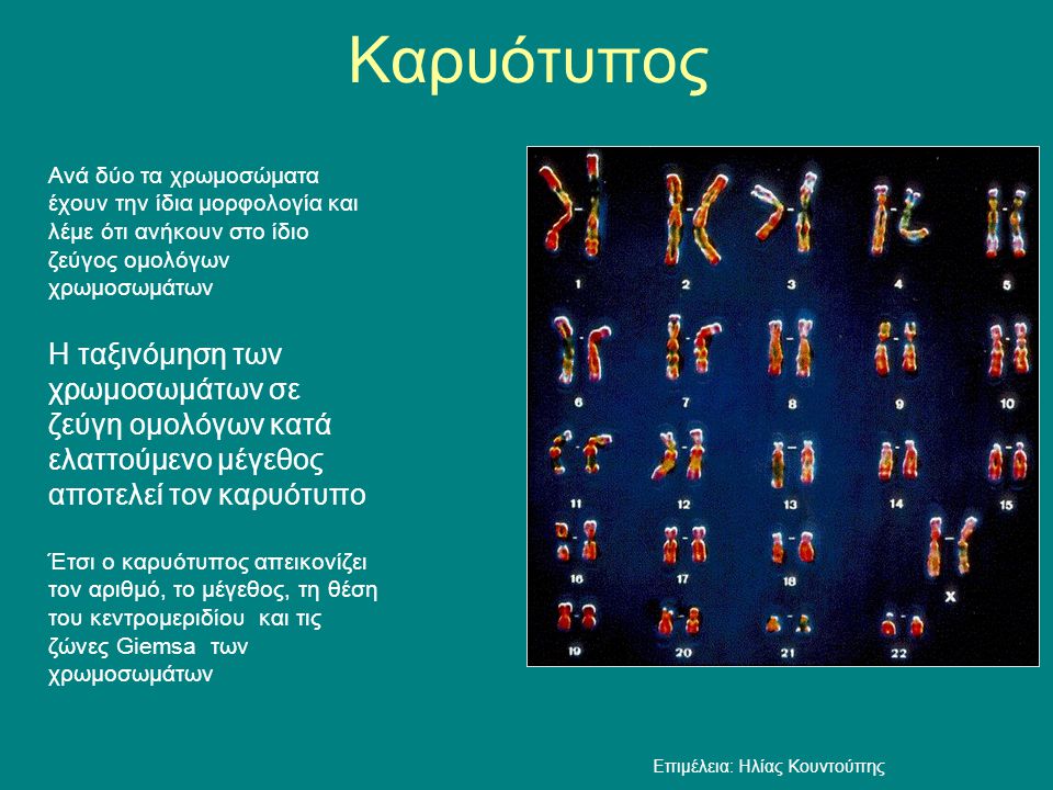
Η οργάνωση του γενετικού υλικού - ppt κατέβασμα

Solved 164. A 24-year-old female presented with an ulcer in | Chegg.com

Restoring production of hippocampal neurons in old age | Nature Neuroscience

Wright-Giemsa stain | VWR

Candida glabrata in Giemsa stain of sputum: Introduction, Pathogenecity

Lymph node biopsy consistent with marginal zone lymphoma (40×,... |  Download Scientific Diagram
Lymph node biopsy consistent with marginal zone lymphoma (40×,... | Download Scientific Diagram

PLOS Medicine: Evaluation of splenic accumulation and colocalization of  immature reticulocytes and Plasmodium vivax in asymptomatic malaria: A  prospective human splenectomy study
PLOS Medicine: Evaluation of splenic accumulation and colocalization of immature reticulocytes and Plasmodium vivax in asymptomatic malaria: A prospective human splenectomy study

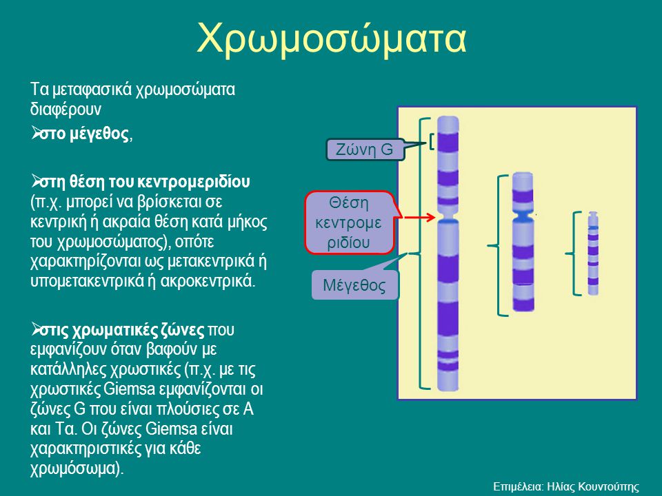
Η οργάνωση του γενετικού υλικού - ppt κατέβασμα

The immune cells by Giemsa stain (A, B) and toluidine blue (C, D). (A,... |  Download Scientific Diagram
The immune cells by Giemsa stain (A, B) and toluidine blue (C, D). (A,... | Download Scientific Diagram

Bone marrow aspiration smear (May-Giemsa staining, ×1,000). Note: The... |  Download Scientific Diagram
Bone marrow aspiration smear (May-Giemsa staining, ×1,000). Note: The... | Download Scientific Diagram
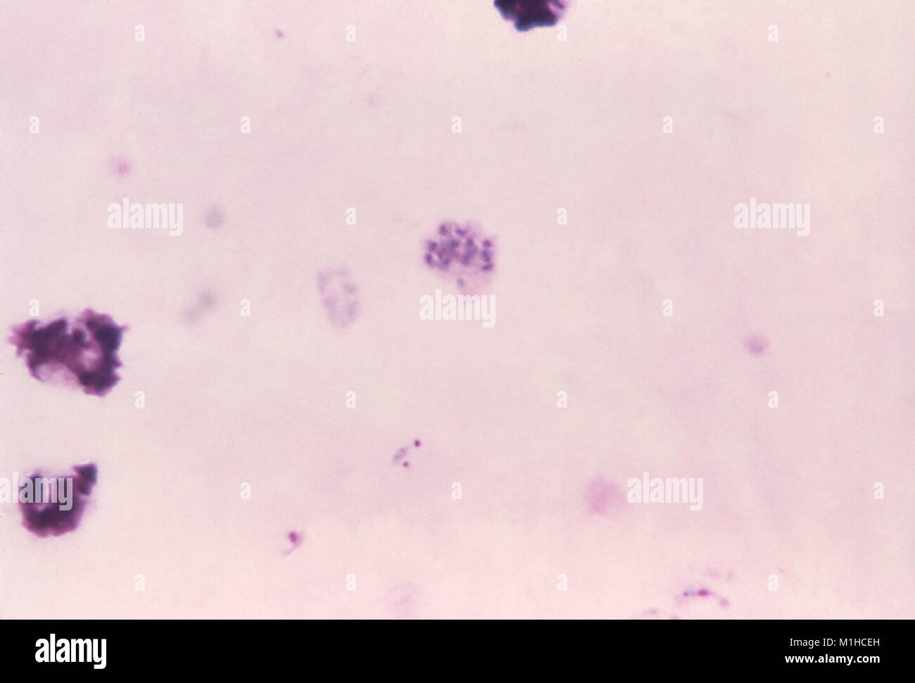
Plasmodium vivax schizont revealed in thick micrograph film using Giemsa  stain, 1971. Image courtesy Centers for Disease Control (CDC) / Dr Mae  Melvin Stock Photo - Alamy
Plasmodium vivax schizont revealed in thick micrograph film using Giemsa stain, 1971. Image courtesy Centers for Disease Control (CDC) / Dr Mae Melvin Stock Photo - Alamy
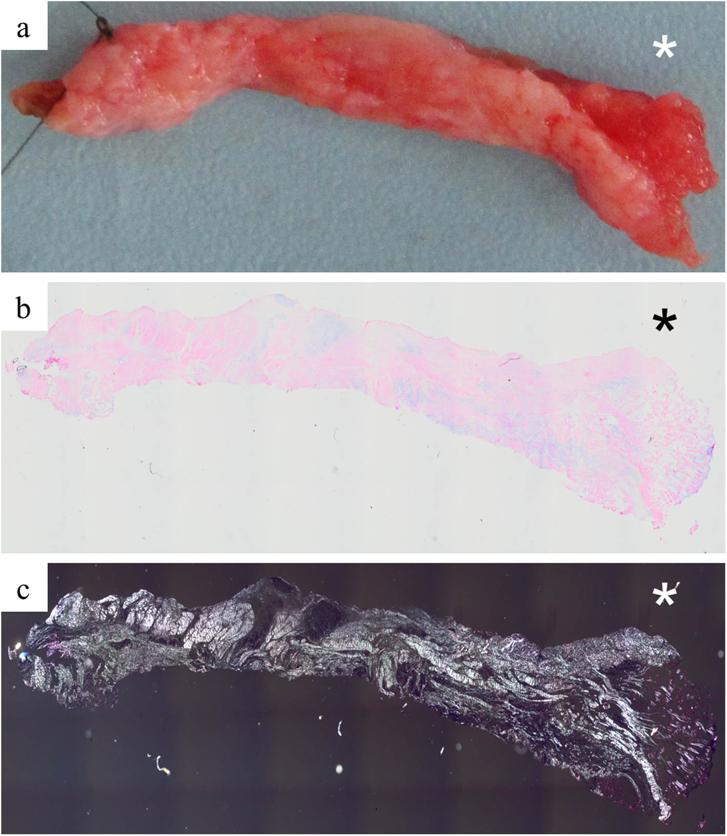
The acromioclavicular ligament shows an early and dynamic healing response  following acute traumatic rupture | BMC Musculoskeletal Disorders | Full  Text
The acromioclavicular ligament shows an early and dynamic healing response following acute traumatic rupture | BMC Musculoskeletal Disorders | Full Text
Blood | Microscopy of Nature

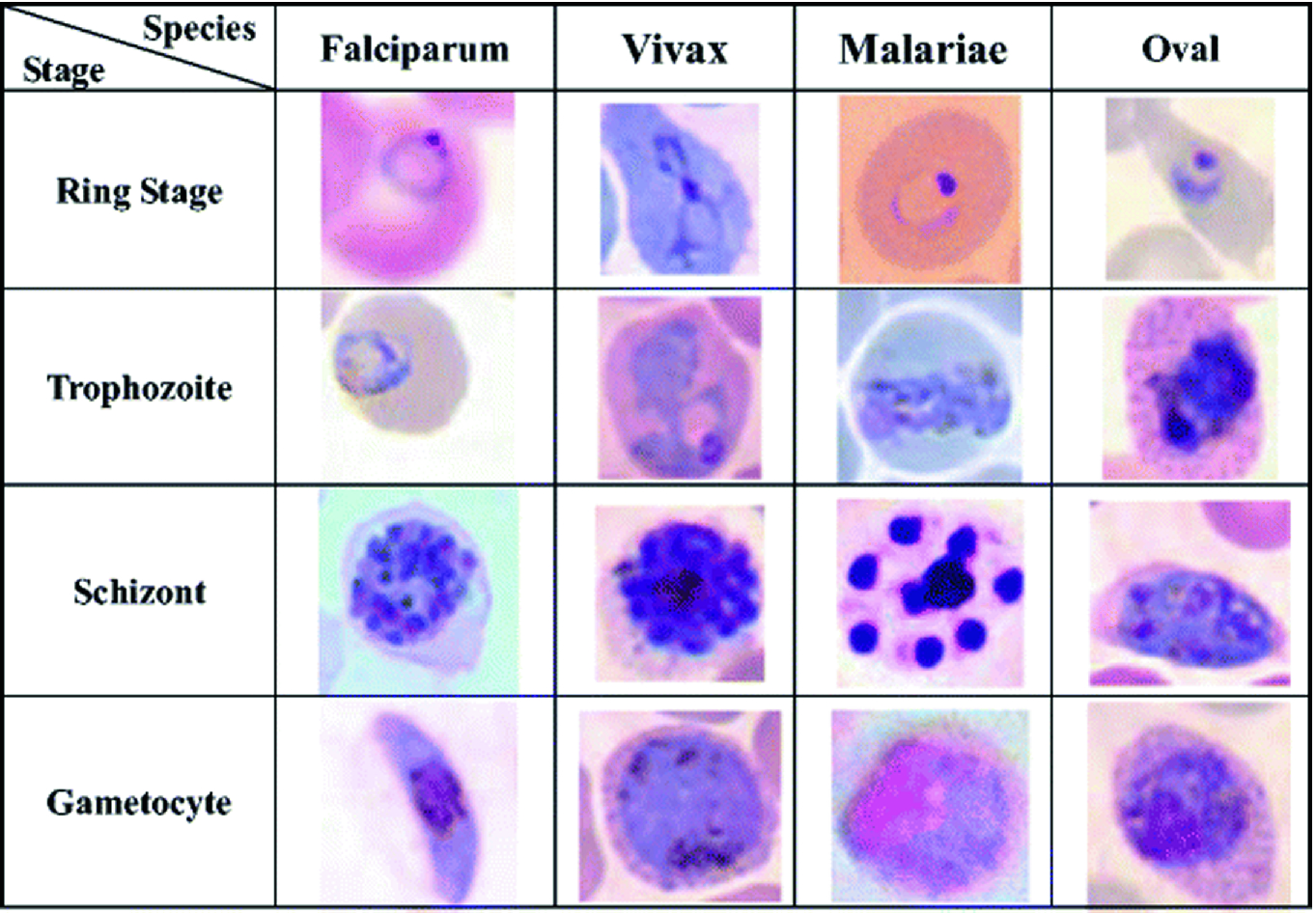
Malaria Detection on Giemsa-Stained Blood Smears Using Deep Learning and  Feature Extraction | SpringerLink
Malaria Detection on Giemsa-Stained Blood Smears Using Deep Learning and Feature Extraction | SpringerLink
Η οργάνωση του γενετικού υλικού - ppt κατέβασμα

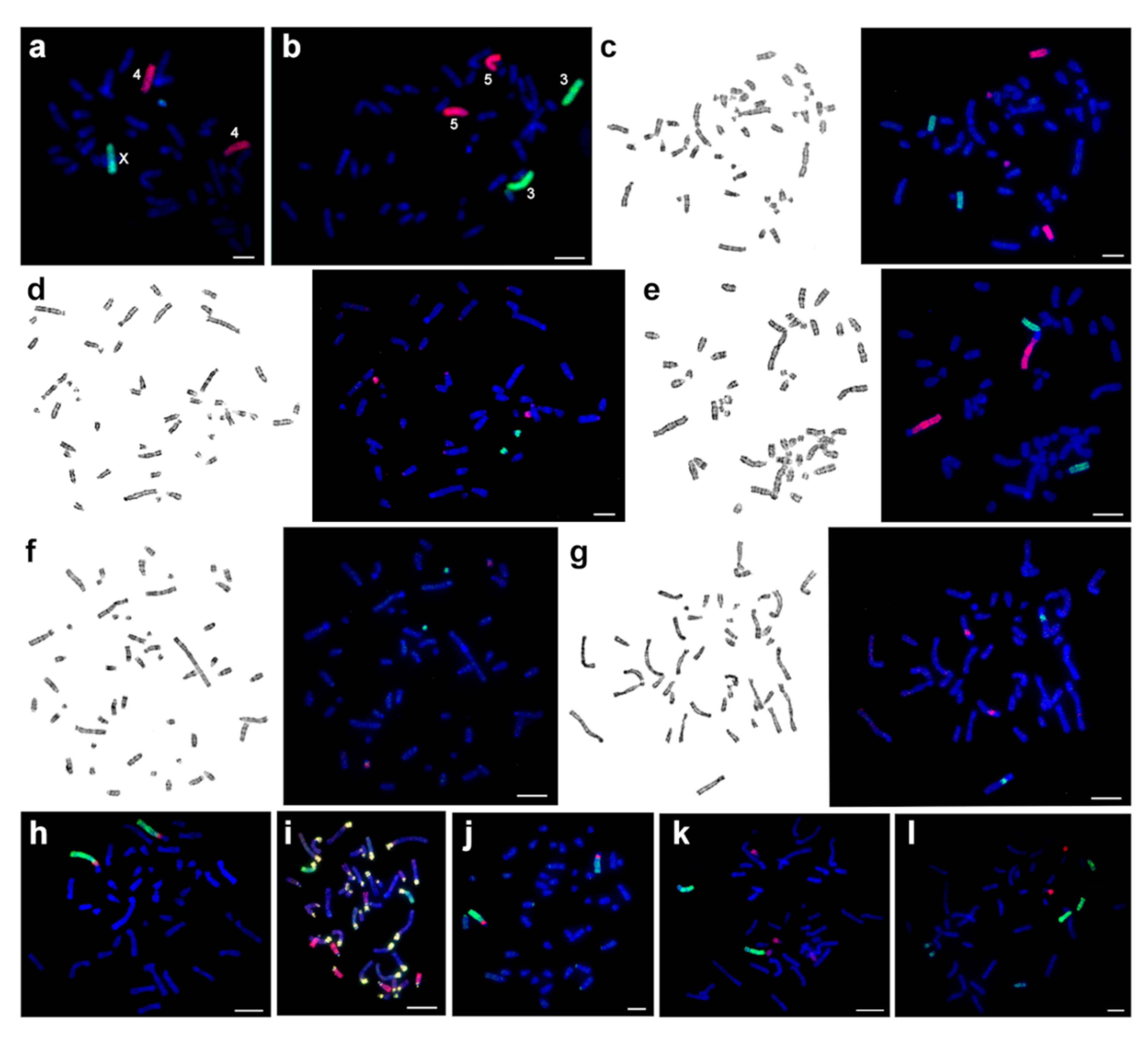
Genes | Free Full-Text | New Data on Comparative Cytogenetics of the  Mouse-Like Hamsters (Calomyscus Thomas, 1905) from Iran and Turkmenistan |  HTML
Genes | Free Full-Text | New Data on Comparative Cytogenetics of the Mouse-Like Hamsters (Calomyscus Thomas, 1905) from Iran and Turkmenistan | HTML
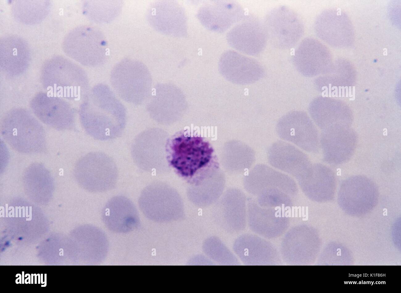
This thin film Giemsa stained micrograph depicts a Plasmodium vivax  microgametocyte. The gametocytes, male (microgametocytes) and female  (macrogametocytes), are ingested by an Anopheles mosquito during its blood  meal. The parasites? multiplication in
This thin film Giemsa stained micrograph depicts a Plasmodium vivax microgametocyte. The gametocytes, male (microgametocytes) and female (macrogametocytes), are ingested by an Anopheles mosquito during its blood meal. The parasites? multiplication in

Figure 5 | A combined banding method that allows the reliable  identification of chromosomes as well as differentiation of AT- and GC-rich  heterochromatin | SpringerLink
Figure 5 | A combined banding method that allows the reliable identification of chromosomes as well as differentiation of AT- and GC-rich heterochromatin | SpringerLink